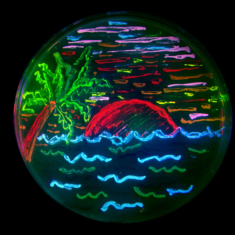
“Three U.S.-based scientists won a Nobel Prize on Wednesday for turning a glowing green protein from jellyfish into a revolutionary way to watch the tiniest details of life within cells and living creatures”: http://apnews.excite.com/article/20081008/D93MK27O0.html
Of course (getting on soapbox), few people give a shit about this stuff, as I have said before. There will be more reporting on OJ Simpson’s sentencing than the Nobel Prizes.
Like, later on, when GFP is no longer the newest phenomenon on the scientific market and is more accessible, could it be used to color certain organelles, proteins, DNA segments, RNA, pathogens, etc on a prepared slide of, let’s say, infected neuroglia cells? I don’t know what method they would utilize; I’m just 16 and curious.
@Sloan. Good questions, all. In fact, pretty much everything that you listed has been done using GFP. Here is a picture of a cell labeled with several versions of GFP to mark different organelles. This picture is of cells labeled with Histone-GFP, thus labeling the nucleus. Mycobacteria, flies, mice, you name it can all be labeled with GFP.
Sure you could. If you are truly interested, have your biology teacher contact a local university for some bacterial strains carrying GFP. If you need more help, send me a PM and maybe we could work something out…
I should add that it might be a tad difficult for your high school to pull off, sadly. As a fluorescent protein, you need special equipment to detect the fluorescence (a special microscope, for example). If your school has that, great! Otherwise, maybe you want to look into spending some time over the summer in a lab at your local university/medical school.